lv wall segments on echo | wall segments echo printable charts lv wall segments on echo The right and the left ventricles are divided into six walls. The left ventricular walls are anterior, anteroseptal, inferoseptal, inferior, inferolateral and anterolateral. The right ventricle has a free . 2M Followers, 1,100 Following, 594 Posts - Emily Miller (@emilyfayemiller) on Instagram: "Podcast: @nowweretalkingbaby (All the fun happens on my tiktok & snapchat) Animal lover Management: @hldtalent"
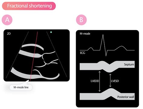
0 · what is fractional shortening echo
1 · wall segments echo printable charts
2 · left ventricular wall segment model
3 · ejection fraction vs fractional shortening
4 · echo wall segments labeled
5 · echo wall motion segments images
6 · echo Lv function
7 · Lv function assessment by echo
Epi leather is a Vuitton-specific creation that’s textured with wavy micro-ridges. The only monogram visible here is a solo LV that is subtly branded into the grain. The house used the glossy.
tricular [LV] size and ejection fraction [EF], left atrial [LA] volume), outcomes data are lacking for many other parameters. Unfortunately, this approach also has limitations.Semi quantitative wall motion score (1-4) can be assigned to each segment to calculate the LV wall motion score index (sum score of all segments assessed / # segments assessed). .
The American Society of Echocardiography (ASE) issued a 16-segment left ventricle model for wall motion assessment. The American Heart Association's (AHA) 17-segment model has an additional apical segment .

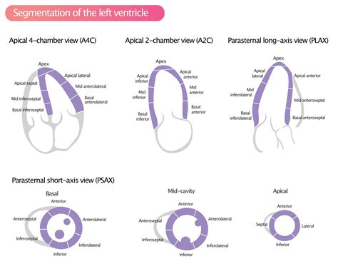
The right and the left ventricles are divided into six walls. The left ventricular walls are anterior, anteroseptal, inferoseptal, inferior, inferolateral and anterolateral. The right ventricle has a free .Regional wall motion can be assessed with a scoring system developed by the American Society for Echocardiography (ASE). The left ventricle is divided into 17 segments (Figure 1) and each .
Identification and classification of left ventricular (LV) regional wall motion (RWM) abnormalities on echocardiograms has fundamental clinical importance for various cardiovascular disease. Calculation of the left ventricular wall motion score index (WMSI) with transthoracic echocardiography allows the semi-quantification of left ventricular ejection .
what is fractional shortening echo
Qualitative estimation of myocardial perfusion contrast echo is inferior to contrast enhanced 2D echocardiography with regard to visibility of all LV segments and appears slightly inferior with .

Physiologic Points in Wall Motion. Transmural extent of infarction is related to regional wall motion. Both acute (6 hr) and subacute (72 hr) 75% thickness infarction moves better than .Standardized myocardial segmentation and nomenclature for echocardiography. The left ventricle is divided into 17 segments for 2D echocardiography. One can identify these segments in multiple views. The basal part is divided into six segments of 60° each.
tricular [LV] size and ejection fraction [EF], left atrial [LA] volume), outcomes data are lacking for many other parameters. Unfortunately, this approach also has limitations.
Semi quantitative wall motion score (1-4) can be assigned to each segment to calculate the LV wall motion score index (sum score of all segments assessed / # segments assessed). Regional Wall Motion during Infarction and Ischemia
wall segments echo printable charts
The American Society of Echocardiography (ASE) issued a 16-segment left ventricle model for wall motion assessment. The American Heart Association's (AHA) 17-segment model has an additional apical segment "cap" added to harmonize left ventricular segment nomenclature with nuclear cardiology and cardiac magnetic resonance imaging.
The right and the left ventricles are divided into six walls. The left ventricular walls are anterior, anteroseptal, inferoseptal, inferior, inferolateral and anterolateral. The right ventricle has a free wall and a septal wall (left ventricular septal wall).Regional wall motion can be assessed with a scoring system developed by the American Society for Echocardiography (ASE). The left ventricle is divided into 17 segments (Figure 1) and each segment should be assessed for contractility (motion) using the following scoring system:
Identification and classification of left ventricular (LV) regional wall motion (RWM) abnormalities on echocardiograms has fundamental clinical importance for various cardiovascular disease. Calculation of the left ventricular wall motion score index (WMSI) with transthoracic echocardiography allows the semi-quantification of left ventricular ejection fraction (LVEF).
Qualitative estimation of myocardial perfusion contrast echo is inferior to contrast enhanced 2D echocardiography with regard to visibility of all LV segments and appears slightly inferior with regards to interobserver agreement (IOA), while both are superior to unenhanced 2D echocardiography.Physiologic Points in Wall Motion. Transmural extent of infarction is related to regional wall motion. Both acute (6 hr) and subacute (72 hr) 75% thickness infarction moves better than 100% thickness. Distribution of wall motion is correspondent to coronary artery supplying the area.
Standardized myocardial segmentation and nomenclature for echocardiography. The left ventricle is divided into 17 segments for 2D echocardiography. One can identify these segments in multiple views. The basal part is divided into six segments of 60° each.tricular [LV] size and ejection fraction [EF], left atrial [LA] volume), outcomes data are lacking for many other parameters. Unfortunately, this approach also has limitations.Semi quantitative wall motion score (1-4) can be assigned to each segment to calculate the LV wall motion score index (sum score of all segments assessed / # segments assessed). Regional Wall Motion during Infarction and Ischemia The American Society of Echocardiography (ASE) issued a 16-segment left ventricle model for wall motion assessment. The American Heart Association's (AHA) 17-segment model has an additional apical segment "cap" added to harmonize left ventricular segment nomenclature with nuclear cardiology and cardiac magnetic resonance imaging.
left ventricular wall segment model
The right and the left ventricles are divided into six walls. The left ventricular walls are anterior, anteroseptal, inferoseptal, inferior, inferolateral and anterolateral. The right ventricle has a free wall and a septal wall (left ventricular septal wall).Regional wall motion can be assessed with a scoring system developed by the American Society for Echocardiography (ASE). The left ventricle is divided into 17 segments (Figure 1) and each segment should be assessed for contractility (motion) using the following scoring system:

Identification and classification of left ventricular (LV) regional wall motion (RWM) abnormalities on echocardiograms has fundamental clinical importance for various cardiovascular disease. Calculation of the left ventricular wall motion score index (WMSI) with transthoracic echocardiography allows the semi-quantification of left ventricular ejection fraction (LVEF).Qualitative estimation of myocardial perfusion contrast echo is inferior to contrast enhanced 2D echocardiography with regard to visibility of all LV segments and appears slightly inferior with regards to interobserver agreement (IOA), while both are superior to unenhanced 2D echocardiography.
gucci sylvie animal studs
gucci the cow
Save time. Automatically transform AJAX requests from JavaScript datatable into LINQ queries. ⚡️. Simple and powerful. Zero configuration for simple cases and powerful .Ēnu diena 2024. pie. laiko. profe. siju. 04/04/2024. Aizmirsi paroli? pieslēgties. reģistrēties. SASNIEGUMIEM IEDVESMO: atbalsta: ĒNU DEVĒJIEM. noderīga informācIja ēnu devējiem. skaitļi. ĒNU Diena skaitļos un faktos. skolēniem. kas jāzina skolēniem par ēnu dienu. partneru piedāvājumi. par ēnu dienu.
lv wall segments on echo|wall segments echo printable charts


